MedVision AI · Medical image archive with an AI assistant
Image, report and AI second opinion in one browser window.
MedVision AI combines study storage and viewing, structured reports for nine specialties, and medical AI models that run on your clinic's own server.
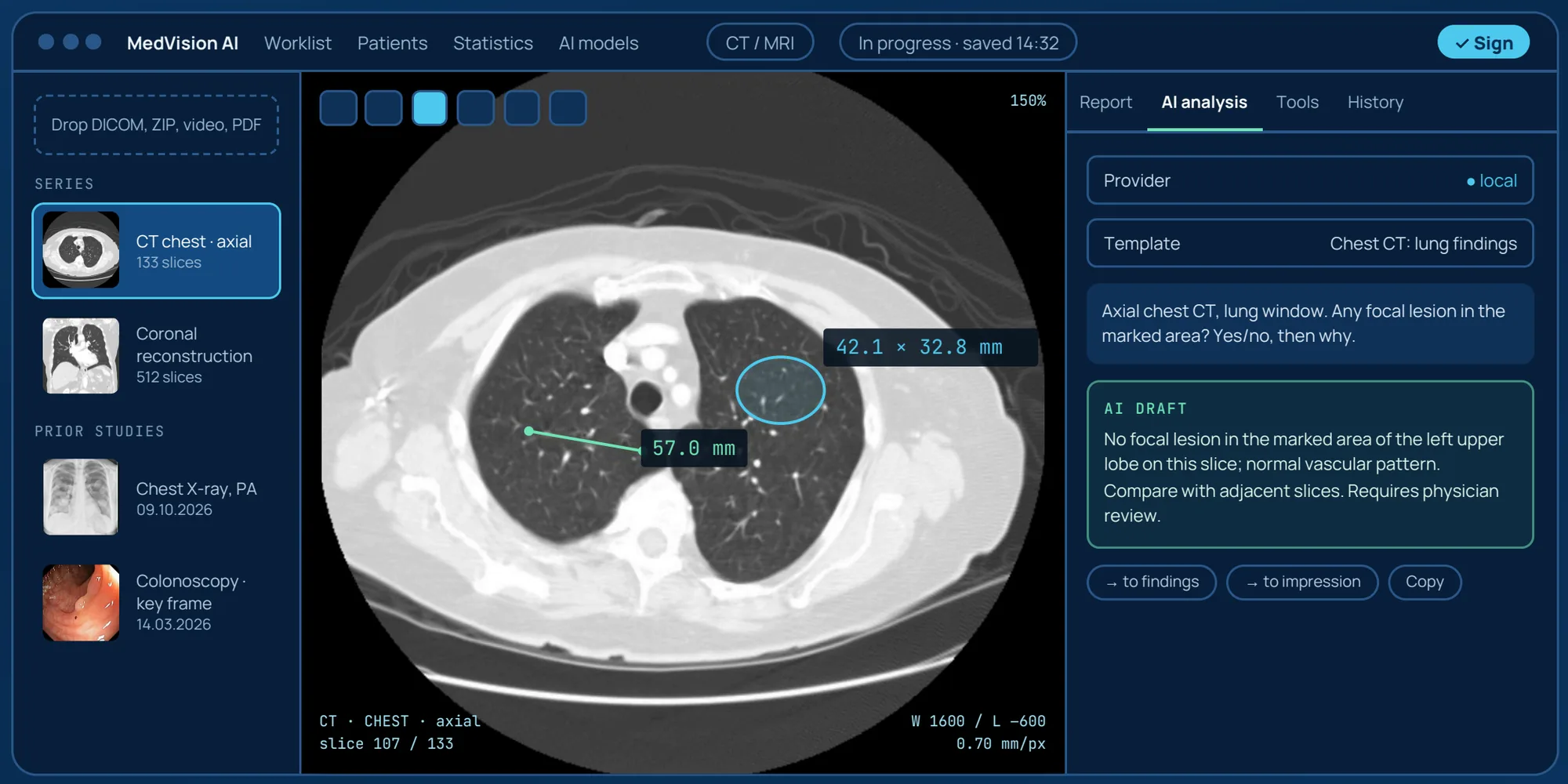
From upload to signature without switching apps
The form turns itself into report text, calculators update as you type, and key images go onto the printed A4 report.
AI runs inside the clinic
All AI models run on your server. Images and reports never leave the hospital network; no cloud services are used.
The physician always decides
The AI answer stays a draft and a second opinion. Signing locks the report, and an audit log records every action and every AI call.
Video
MedVision AI in 53 seconds
A short overview of what MedVision AI does: archive, viewer, local AI and the physician's final say.
Screens
Inside MedVision AI
Interface panels with demo studies: segmentation with volumes, a structured report and the X-ray classifier.
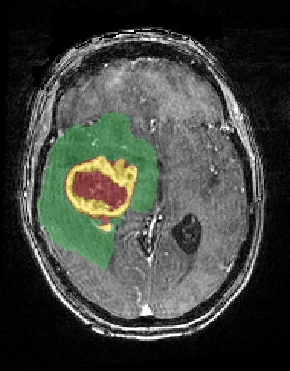 MR brain · T1 post-contrast · slice 111 / 192
| Structure | Volume, ml |
|---|---|
| Whole tumour (WT) | 238.3 |
| Peritumoral oedema | 190.7 |
| Tumour core (TC) | 47.5 |
| Enhancing tumour | 28.8 |
| Necrotic / non-enhancing core | 18.8 |
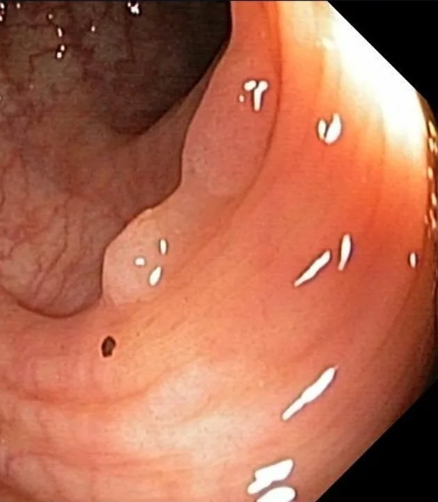 Key frame
| Physician | n | ADR | CIR | Withdr. |
|---|---|---|---|---|
| A (demo) | 35 | 37.1% | 97.1% | 9.3 min |
| B (demo) | 34 | 14.7% | 76.5% | 6.4 min |
| ESGE target | ≥ 25% | ≥ 90% | ≥ 6 min |
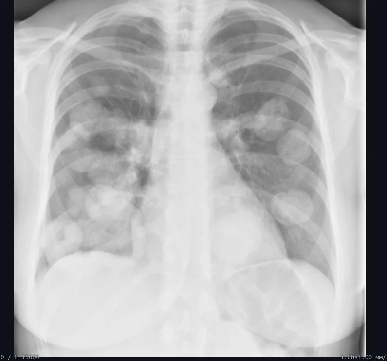
Frontal (PA/AP) chest X-ray only · ≥ 0.6 worth a closer look · around 0.5 is noise
Images: UPENN-GBM (TCIA, CC BY 4.0), HyperKvasir (CC BY 4.0). Patient data and values are from the demo set.
01 — Base product
The complete diagnostic workstation
Archive, viewer, reporting and AI share one screen and one patient record, so nobody copies text between programs.
Browser-based viewer
CT, MRI, X-ray, ultrasound, endoscopy. Three planes, CT window presets, cine loop, ruler and ellipse in mm, annotations on slices.
Import without manual entry
DICOM, ZIP, NIfTI, photos, video and PDF, up to 2 GB per upload. Patients and studies are created from file headers.
Structured reports
Specialty forms with lists of lesions, polyps and stones. Prostate volume, PSA density, IPSS and BBPS are calculated automatically.
AI image analysis
Medical AI models on your server. About 200 ready-made question templates; one click sends the answer to the findings.
Report assistant
Drafts the impression and recommendations, checks the text for contradictions and explains the result to the patient in plain language.
Numbers, not guesses
A classifier for 18 findings on frontal chest X-rays, and CT/MRI segmentation with volumes and density in HU.
Video and key frames
Colonoscopy and cystoscopy recordings play in the browser. One click adds a frame with a finding to the report.
Quality statistics
ADR, PDR, caecal intubation rate and withdrawal time for each physician. Workload by specialty for any period.
02 — Study workflow
Five steps from file to signed report
The AI is one step in the middle, not the end of the process. The physician reviews, edits and signs.
- 01
Upload
Drop a folder or ZIP. Hundreds of files group into series; the patient is matched by ID, name and date of birth.
- 02
Review
Scroll slices, change window and plane, measure with ruler and ellipse, add labelled arrows.
- 03
Report
Fill in the specialty form. “Findings from fields” turns it into coherent text.
- 04 · AI
Second opinion
Select a series and a question template. The answer streams in and is saved to history.
- 05
Sign and print
Signing records the physician and time. An A4 report with your letterhead and key images goes to print or PDF.
03 — Reports and AI
Clinical scales built into the forms
Each specialty has its own form. Totals and volumes are calculated as you type.
| Specialty | Sections and classifications | Calculated automatically |
|---|---|---|
| Urology | Kidneys (SFU, Bosniak), stones with size and HU, bladder, prostate (PI-RADS v2.1, lesions by zone), scrotum (varicocele, Dubin), cystoscopy, uroflowmetry | Prostate and bladder volume, PSA density, testicular volume, IPSS total with interpretation |
| Colonoscopy | Bowel prep (BBPS); polyps: Paris, NICE, JNET, Kudo, resection method, histology; IBD: Mayo, UCEIS, SES-CD, Rutgeerts | BBPS total with adequacy, UCEIS total |
| Upper GI | Oesophagitis (LA), Barrett's (Prague C/M), varices, atrophy (Kimura–Takemoto), H. pylori, ulcers (Forrest) | — |
| CT / MRI | Technique, lesions with follow-up, Lung-RADS, LI-RADS, PI-RADS, Bosniak, O-RADS MRI | Structure volumes via segmentation |
| Radiography | Projections, quality, chest, mammography (ACR breast density, BI-RADS) | Scores for 18 findings from the classifier |
| Ultrasound | Abdomen, thyroid (TI-RADS), breast (BI-RADS) | Thyroid lobe volume |
| Gynaecology | Uterus, endometrium, fibroids (FIGO), ovaries (O-RADS), colposcopy (IFCPC 2011) | — |
| Dermatology | Lesion, ABCD rule (Stolz), 7-point checklist | TDS and checklist total with interpretation |
| Ophthalmology | Diabetic retinopathy OD/OS, macular oedema, retinal thickness on OCT | — |
04 — Local AI
Three independent sources of a second opinion
Language models describe; specialised models measure. When they disagree, the numbers win and the physician decides.
Local · primary
Primary medical model
X-ray, CT and MRI slice sets, endoscopy, ultrasound, dermoscopy. Fast, concise answers to the physician's targeted questions.
Local · cross-check
Second independent model
An independent second opinion and structured reports. Agreement between the two models increases confidence in the result.
Numbers
Specialised models for verification
Chest X-ray classifier: independent scores for 18 findings on frontal (PA/AP) chest X-rays in 10–30 seconds. Segmentation: 117 structures on CT, 50 on MRI, colour masks, volume in ml and density in HU.
Formats
Accepts what your department has
| Format | What happens |
|---|---|
| DICOM, ZIP | Grouped into series, one thumbnail per series |
| NIfTI | Volume in radiological orientation |
| JPG, PNG, TIFF | Endoscopy, dermoscopy, fundus, photos |
| MP4, MOV, AVI, MKV | Plays in the browser, automatic transcoding |
| Referrals and histology next to the images |
Colonoscopy quality
Indicators for each physician
| Indicator | What it measures | Target |
|---|---|---|
| ADR | Share of studies with an adenoma on histology | ≥ 25% |
| CIR | Examination to the caecal pole | ≥ 90% |
| Withdrawal | Mean time and share ≥ 6 minutes | ≥ 6 min |
| Bowel prep | BBPS ≥ 6 and ≥ 2 in each segment | ≥ 90% |
| PDR, SDR | Polyps and serrated lesions | — |
Targets follow the ESGE performance measures for colonoscopy. ADR is reliable only once histology is entered for each polyp.
Privacy
What the local AI model on your server receives
Pixels of selected slices and the physician's question
Age, sex, indication, study type
Technical tags: modality, series, slice thickness
Report text, without images
Name, ID, date of birth, study dates, insurance numbers
05 — Customisation and deployment
Adapts to your clinic
The base product is ready to use. On top of it we add features for each institution's needs, without limits.
- PACS integrationDICOMweb, studies received directly from modalities
- HIS and LIS connectionHL7 and FHIR: orders, results, histology
- Single sign-onAD, LDAP, SSO, HTTPS and air-gapped networks
- New specialtiesYour institution's forms, scales and print templates
- Voice inputDictation of findings and impression
- Model selection and fine-tuningQuality validated on your data
- Follow-up comparisonAutomatic report on current and prior studies
- Branches and consultsShared archive and remote second opinion
Who it is for
One tool for many departments
- Radiology. CT, MRI, X-ray, ultrasound: series archive, measurements, segmentation with volumes and a structured report in one window.
- Endoscopy and urology. Key frames from recordings, forms with Paris, PI-RADS and IPSS, quality indicators per physician.
- Private clinics. A fast start without a PACS: one server, browser access, reports with your letterhead and clear explanations for patients.
Deployment
No installation on workstations
- The server sits in the hospital network; physicians work in a standard browser.
- The database and files live in one folder, so a backup is a simple copy.
- Local AI models run on the institution's server. We help define the hardware requirements.
- Three roles: administrator, physician, viewer. The log records sign-ins, views, edits, signatures and AI calls.
- Getting started · 01
Demo, 30 minutes
We show the full path from study upload to signed report.
- 02
Pilot on your server
We deploy the system in your network; physicians work with their own studies.
- 03
Rollout and customisation
We put the base product into service, add the features you need and train your staff.
Request a demo
See MedVision AI on your cases.
We'll agree a time and discuss what your department needs.
The AI models in the system are not medical devices: their output is a draft and a second opinion, and the physician writes and signs the conclusion. “On request” features are developed to customer requirements. Interface illustration uses fictional patient data.